A Radiologist’s Approach to TBI Case Evaluation – Dr. David Levey


“What I’m looking for is: What is the correct diagnosis? What happened to this patient? Is he or she being treated correctly or properly? And that’s a concern of mine.” – Dr. David Levey
Welcome to Winning The TBI Case, hosted by trial lawyer Tom Crosley. Listen as Tom provides practical, powerful guidance for litigating traumatic brain injury cases. This podcast is meant for litigation attorneys who are ready to explore the essentials of TBI cases to confidently represent clients who have suffered traumatic brain injuries.
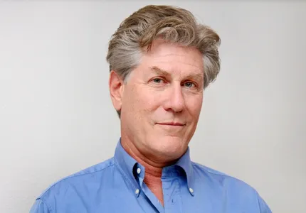
Tom is joined by Dr. David Levey, a diagnostic radiologist and forensic neuroradiology expert who evaluates traumatic brain injury cases for both plaintiffs and the defense. Dr. Levey walks through the detailed process radiologists use to evaluate imaging, medical records, symptoms, and clinical history when analyzing TBI cases in both medical and litigation settings.
What’s In This Episode
Dr. David Levey explains how radiologists evaluate traumatic brain injury cases and how imaging findings fit within the broader clinical and litigation picture. He also shares practical insight into the role imaging plays in TBI litigation and best practices for trial lawyers working with radiology experts.
Key Insights for Trial Lawyers
- Radiologists Evaluate More Than Images: Forensic radiology involves an extensive analysis of imaging combined with medical records, symptoms, clinical history, and causation evidence in context.
- “Normal” Scans and Imaging Do Not Rule Out TBI: There are times when traumatic brain injuries will not appear on routine CT imaging. MRI studies can reveal subtle abnormalities not visible on CT scans. Advanced imaging such as DTI, tractography, and volumetric imaging can provide additional detail and should always be considered alongside symptoms, testing, and functional changes.
- Effective TBI Litigation Requires Collaboration: Radiologists often work closely with attorneys and other experts to help evaluate complex brain injury cases.
Dr. Levey pulls back the curtain on the detailed analytical process radiologists use to evaluate brain injury cases in both medical and forensic settings and shares how he works with both plaintiff and defense teams.
Preparation Matters
This episode is for trial lawyers who want a clearer understanding of how radiologists evaluate traumatic brain injury cases and how imaging evidence fits into litigation strategy. If you’ve ever struggled with “normal imaging” defenses or wanted insight into how to best leverage your radiology experts in TBI cases, then you’ve come to the right place.
Referenced and Mentioned
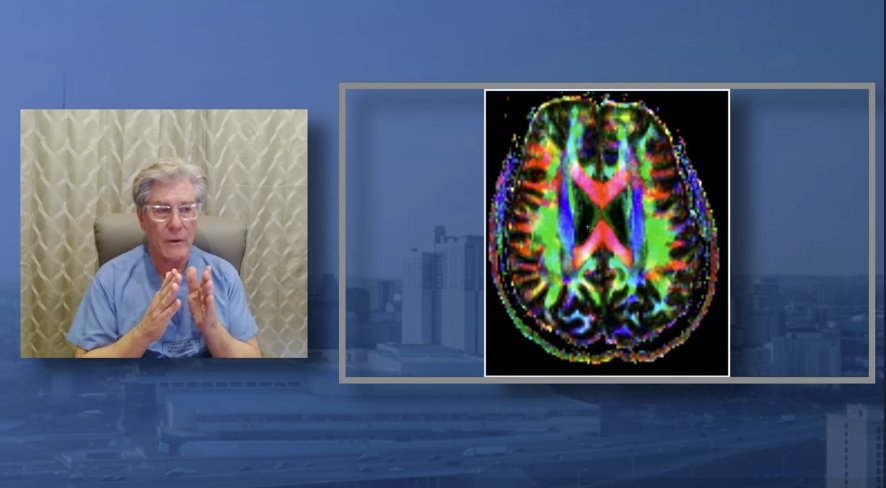
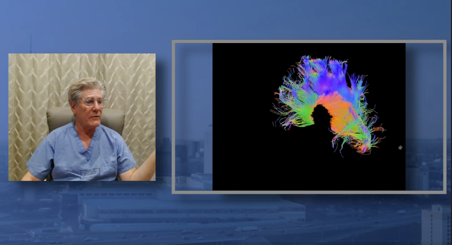
Dr. Levey and Tom reference tractography imaging beginning at time code 22:49 which can be seen on the video version of the podcast. Screenshots below for our audio listeners.
Connect
- Connect directly with Dr. David Levey by visiting: www.expertradiologist.org
- Subscribe to Winning The TBI Case on Apple Podcasts, Spotify, YouTube or wherever you get your podcasts.
- Follow on Facebook, LinkedIn, and Instagram
- And remember, don’t just listen to the strategies, implement them!
Winning the TBI Case — A Radiologist’s Approach to TBI Case Evaluation
Host: Tom Crosley Guest: Dr. David Levey Runtime: 37:08
[00:00] Dr. David Levey (cold open): Follow-up MRIs can be valuable, particularly in two cases. One, if you have found nothing abnormal on imaging structurally, right? Yet the patient is so abnormal. The family doesn’t recognize them anymore. Work’s not going well. The tasks they used to do, they can’t do them anymore. Well, you know something’s wrong.
[00:20] Announcer: TBI cases can feel like navigating uncharted territory. Your client’s injury is real, but proving it — that’s where the real challenge begins. If you’ve ever felt like you’re missing crucial pieces of the puzzle, you’re in the right place. Welcome to Winning the TBI Case, where Tom Crosley shares the insights he’s gained from over 60 injury trials. Not just the victories, but the lessons learned along the way that make all the difference. Winning the TBI Case: strategy, science, and storytelling.
[00:55] Tom Crosley: Welcome back to Winning the TBI Case. I’m Tom Crosley, and today we’re gonna have an exciting episode. We’re gonna be talking about one of the most misunderstood areas in traumatic brain injury litigation, and that is brain imaging. Our guest today is Dr. David Levey, a diagnostic radiologist with a clinical practice reading brains and spines, and he also happens to serve as an expert witness from time to time in legal cases. About half of his work is on the defense side; about half is on the plaintiff side. So he’s one of those expert witnesses who is exactly what we want: science first, advocacy second. Somebody who does both sides and calls the balls and strikes like he sees them.
I wanted to start with a little bit about you and your background. What led you to go to medical school? How did you pick radiology as your specialty? Kind of bring us current to modern times.
[01:59] Dr. David Levey: Well, when I was 11, I read a book called The Defense Never Rests, and that was it. I was going to be a lawyer. And in the next few years, that evolved. I went to college, got two different bachelor’s degrees, really heading towards the neurosciences, and a master’s degree at UCLA before medical school. Then after medical school: internship, a trauma internship, four years of radiology at USC, and a fellowship. After 17 years coming out of the machine, I finally got my first job doing just that — neuroradiology, musculoskeletal imaging — here in Texas. That was the first job, and here I am.
[02:45] Tom Crosley: Tell us about how forensic neuroradiology — for example, in a brain injury case — differs from day-to-day clinical neuroradiology.
[02:53] Dr. David Levey: It’s extraordinary, the difference. I didn’t know about it until I started this 15 years ago. If you’re reading an MRI of the brain as a radiologist, you get the case; the case comes up. You get some clinical data. You may get some clinical information faxed to you. Some of it’s sketchy, some of it’s from the patient. You may get some nursing notes, from the PA or the doctor, but it’s variable. If you get 10 pages of notes, you’re really doing great. If you get a page or two, that’s acceptable too. Many of the diagnoses are fairly complete, and others may have “headache” or “head pain” or “trauma” — no date, no description, no time. You don’t have any idea.
So you are absolutely starting from the beginning and working your way through the brain MRI and trying to get that diagnosis. So you’re looking for everything under the sun. You can’t focus on trauma or anything. And that takes you X amount of time. You know, a brain MRI may take two minutes or two hours. I’ve had one take two days — spent two days on it, getting through it, it was so complicated. But it’s a finite experience.
But forensic radiology — you get reams of medical records and a number of opinions from everywhere. From the police report at the scene of the accident, all the way through the ER and the ICU and the consultants and surgery or not. Then the patient finally is released from the hospital and has all these follow-up appointments with many other experienced, seasoned experts: pain people, clinical psychology people, PM&R, et cetera. And the vast amount of information I can use to put a story together is what I think attracted me at first to this field. All the imaging available plus all the medical records together allows me to tell a story — not only with the radiology and the imaging, but the medical process and the path the patient took as well.
[04:57] Tom Crosley: Some radiologists that serve as expert witnesses don’t like to look at medical records. They just want to look at the imaging and dictate a report based on what they see. But it sounds like you like to look at the whole picture, including medical records. Why is that?
[05:10] Dr. David Levey: I think it’s important. I do like it, you’re right. But I also think it’s important. I think you’re missing something if you’re not doing that, because, first of all, from the correct diagnosis flows the correct treatment. I want to get involved with any patient — it doesn’t matter, defense, plaintiff, or whatever the situation is. What I’m looking for is: What is the correct diagnosis? What happened to this patient? And is he or she being treated correctly or properly? And that’s a concern of mine.
Because I see that if the radiologist who read the primary studies got it wrong, the whole crowd — medical, surgical, everybody — they’re following down the wrong pathway. And that’s the classic GIGO I see all the time: garbage in, garbage out. You get the wrong diagnosis, and there’s surgery in the wrong place, or no surgery where he needs it, or treatment that is inappropriate because the diagnosis was not made correctly in the beginning. So medical records really help with that. You hardly ever get that when you just get a case, read it, get a case, read it, like in a radiology practice.
[06:15] Tom Crosley: It sounded like your explanation of the difference between clinical radiology and forensic radiology was that sometimes, clinically, you might only spend a few minutes reading a brain scan, but if it’s forensic — meaning it’s going to be used in a court case — then you’re going to spend probably a lot more time. Is that what I’m hearing?
[06:35] Dr. David Levey: I do, and I see them over and over again, because it may take weeks to get the consultative report out. But then if you have a deposition, or are called upon in this case for any other reason, it takes weeks, maybe months, that you’re going over and over these things. I currently have a case that’s in its sixth year.
[06:55] Tom Crosley: Let’s take a step back, Dr. Levey, and have you tell us what radiologists typically do in evaluating brain injury. And I’m not talking about experts; I’m talking about clinical radiologists. What’s their role?
[07:07] Dr. David Levey: You get the scan — you pull it up on your reading station. You have a dictaphone, one you might use such as this one; if you’re on YouTube, you can see my dictaphone. We live and die by it. And you evaluate the case as you go through hundreds of images.
Now, they’re set up in a reading situation where they’re called sequences, and you’ll do different sequences that weight the brain tissue in different ways. And so you can look at them, if you would, from different angles. If you’re doing a CT scan, it’s very simple. Usually it’s axial images. It’s an X-ray study, and it’s black and white. You kind of zip right through the head axially. But now we can reconstruct these in other planes and see a lot more than we used to. So CT is valuable in the acute setting especially.
But then MRI can have hundreds, and in some cases thousands, of images, and we’re no longer looking at pixels. This is one big thing that has changed. It was pixel imaging for decades in radiology. Like X and Y, a box. And these boxes had images, and these images allowed you to look at the brain. But today we’re looking at voxels — a volume pixel. So you have an X, a Y, and a Z: a width, a height, and a depth in these studies. So now we have contiguous imaging, and we don’t miss a thing. We’re right on top and overlapping as you come through the brain parenchyma itself.
And in doing so, you get the base of the brain, you get the orbits, you get the sinuses, the inner ear, the cerebellum, the supratentorial compartment. It’s a fallacy that you’re just looking at the brain only. You look at everything that’s in the field of view. You’re responsible for that. As a diagnostic radiologist, to answer your question, you get that study, you bang through those images, you make your report, you make sure you don’t miss anything, and you’re on to the next one.
[09:10] Tom Crosley: In a clinical setting, what is the typical amount of time that a radiologist will look at a brain scan?
Dr. David Levey: It’s minutes. It’s not hours, that’s for sure.
Tom Crosley: And if it’s minutes in a clinical setting — because of the volume of the practice and the fact that they’ve got to read 50 or 60 or more scans a day — what is the amount of time that you typically spend reading that same MRI scan if it’s a litigation case and you’ve been retained as an expert for the defense or the plaintiff?
[09:37] Dr. David Levey: First off, it has to be a little longer, because I have someone else’s report and I have the images. So I’m going to go through the images, and I’m going to be looking to see what the primary radiologist — I call them the primary radiologist; you can call it the treating radiologist — what they said about the study and what they didn’t. Whatever’s on the image, you must report. We are not of a standing where we can pick and choose. If it’s an abnormality or a finding and it’s on the scan, you need to report it. And I see a lot of things being left out or misinterpreted.
So my first way through is to see what the radiologist said. The next one is to compare it to any of the other prior examinations. And this is very special in my practice: I have a bird’s-eye view into all the other prior scans that many times the radiologists do not. I amass them, right? We download them; we get everything ready. Whereas that radiologist may have one study — reads that study, and he’s on to the next one. He doesn’t have the advantage of having prior studies. And it is so, so nice. So I have an unfair advantage, and now I can see multiple years, sometimes, of brain scans and then see the current one. That’s important.
So yes, it takes longer, because there’s more material there, and I’m kind of doing my due diligence and a little bit of detective work as well to make sure that all the details have been seen.
[11:01] Tom Crosley: Dr. Levey, in my litigation practice over the last few decades, I’ve had cases — certainly very commonly — where there’s a CT scan done in the ER. Very commonly, it’s read as normal unless there was a fracture or bleeding. Does a normal CT scan rule out a brain injury?
Dr. David Levey: It does not.
Tom Crosley: And is that something that every radiologist in the country should agree upon, or is that up for debate?
[11:24] Dr. David Levey: I think it’s really not up for debate. And it doesn’t matter what side you’re on, or even what specialty you are. I mean, if you look at a brain CT and you don’t see an abnormality, you know that there can be something there. And so many of the ones that are read as “no acute process” — you see these, “no specific abnormality” — those are all true statements, but that doesn’t mean there isn’t something present that’s not available to be visualized on the CT scan.
[11:56] Tom Crosley: When a patient comes in with a suspected head injury and a CT is run on them in the ER, is the main thing they’re looking for fracture or bleeding?
[12:06] Dr. David Levey: Those are the two main things they’re looking for as well. You can also have post-traumatic stroke. You can have edema in the brain — so no blood at all, but a real good contusion can give you edema, and that edema should show up quite rapidly and be seen on a CT. But you’re right: fractures and blood are the number one and number two things, because they’re seen so well. They’re radiodense, and they’re gonna be seen as white on your image.
[12:36] Tom Crosley: What I typically see with MRIs is, if a patient is still symptomatic and weeks have passed by, then an MRI scan is ordered. And often in our mild cases, that one will come back as normal. But is that also just a structural test? It’s looking at the structure of the brain and not the function of the brain?
[12:55] Dr. David Levey: It certainly does look at the structure, but the one thing that it has over CT, or much of the other neuro testing you can do, is that we have structural resolution and contrast resolution. Those are the two big things. MRI can look at the contrast between bone and fluid, fluid and fat, cortex and white matter, white matter and ventricles — all the different things. Vessels: flow and no flow.
It’s structural for sure, but it isn’t as good as CT. It will never be as clear as a CT in terms of anatomical, but it’s okay, because it has gotten so good now. But it also has contrast resolution, which is the most valuable thing that an MRI can provide. And because that relates to blood flow as well — tissue viability, or the absence of viable tissue, and tissues that can be injured; blood that’s old, subacute, or acute — all those things can be seen on the MRI. So it’s just a more versatile test than the CT scan.
[13:55] Tom Crosley: In a plaintiff case, if you are retained early enough to where you can make suggestions about what type of imaging plaintiffs should receive, what suggestions do you typically make?
[14:04] Dr. David Levey: If it’s a trauma case, I typically do not need contrast — that’s IV contrast material injected at the time of the examination. So: a non-contrast 3T MRI of the brain. And I have a specific protocol that covers all kinds of disease processes — both the ones we’re looking for that are trauma-related and totally non-trauma-related — because you have to examine the patient’s brain in totality. You get surprised sometimes by what may pop up.
Would I add a DTI to that? Well, you can’t lose in doing that. And what I mean by that is, the patient’s in the magnet, they’re on the scanner, the tech is doing the study, they’re almost done. All they have to add is three or four more sequences. So if the patient can lie still for another 10 to 20 minutes, depending, and you can acquire this data, then I would say do that as well.
And I would say to do that because, thus far in my practice, I never stop with a DTI — get that study, look at it, report it out, and go. I always take the raw images and send them off to be evaluated for quantitative evaluation. You get this one-two punch, and now you have a ton of data on one brain, on one person. And you may or may not need it, but you certainly have it at your disposal to evaluate.
[15:24] Tom Crosley: And is there a separate sequence that’s run for susceptibility-weighted imaging, or SWI?
[15:29] Dr. David Levey: If we’re saying we’re going to do a DTI, then I’d need to be more specific. I’d package it like this: I want a 3T MRI of the brain, non-contrast, with all the typical protocols utilized, and also those that look for trauma-related things, like an SWI or a GRE. I want the planes that I need, and I also add a number of sequences on the back end for DTI. That in and of itself would be one study that could be very useful, because you could then ship the DTI and brain examination off for quantitative study. So what I like to say is what we’ve done here: we’ve done a 3T non-contrast brain, and we’ve done qualitative evaluation of the brain imaging, and quantitative.
[16:23] Tom Crosley: Is it difficult to get patients access to 3T scanners for brain scans?
[16:27] Dr. David Levey: In North America, not really. In a normal setting, a physician is sending a patient for a study, and he wants the very best type of MRI, he should be sent to an MRI center. We’ve already called them and said, “Hey, you guys have a 3T scanner? Will you always have that available? Oh, you haven’t gotten it yet. I see you’re planning. Thank you.” Click. Go to the next guy.
[16:49] Tom Crosley: So I’ve heard of a 7T scanner as well. When is it appropriate to insist upon a 7T machine?
[16:57] Dr. David Levey: Never, unless you’re doing research on animals. We do not yet know that there is a clinically significant difference in image quality or acquisition on a 3T versus a 7T.
[17:08] Tom Crosley: So if it were your family member who had suffered head trauma and needed an MRI, you’d be recommending a 3T machine for the brain MRI?
[17:17] Dr. David Levey: Yes. But if I knew the 1.5 Tesla, and I knew the tech was good and it was a well-kept, good magnet, it would be just as well to get a 1.5.
[17:28] Tom Crosley: So, Doctor, I had a question about when is the best time to get an MRI. And over the life of a case, which can sometimes go two to three years or even longer, how frequently should the plaintiff be imaged to look for changes over time?
[17:42] Dr. David Levey: If the patient in question has had a trauma, and they’ve gone to the ER and gotten their head CT. If they’re stable, there is nothing holding one back in the first week from getting an MRI, because if the head CT is read as normal, the MRI could still be flagrantly abnormal. And you’d want to know that, because then you could track it over time as you reexamine them over the months or years.
If the head CT is read as abnormal, you could further characterize those abnormalities on MRI so much better than on a CT scan. And getting that detail also allows you not only to tell what’s wrong with the patient now, but, importantly, to prognosticate what may be expected in the future to happen to that brain — and therefore what happens to that person and their ability to think, executive functions and the like, memory, emotions, et cetera. So getting an MRI soon or early in the case — I think a lot of lawyers wring their hands over that. I wouldn’t. I would just go ahead and get it if it’s possible.
[18:50] Tom Crosley: And is it appropriate to order a repeat scan at some point?
[18:53] Dr. David Levey: At some point, I think so. Follow-up MRIs can be valuable, particularly in two cases. One, if you have found nothing abnormal on imaging structurally, right? Yet the patient is so abnormal. The family doesn’t recognize them anymore. Work is not going well. Tasks they used to do, they can’t do them anymore. Well, you know something’s wrong. You’ve had normal brain scans; you definitely want a follow-up MRI, and at high field strength at that.
But if you had a prior scan and it’s abnormal — same thing. You want to track that abnormality, because — and this is the most valuable part — once you have established that the brain has been injured (and so many different injuries can occur), then you want to know what the secondary and tertiary effects on the brain are. After the brain bleeds, or has been injured, or has had edema or infarct, it usually walks through a progression — not linear, a nonlinear progression — to either encephalomalacia, or gliosis, or brain atrophy, and any apoptosis of the actual neurons themselves, giving you groups of dead neuronal tissue that will never come back. That’s all part of the process of figuring out how will this patient do in the future.
[20:10] Tom Crosley: I tried a case last year in another state, and the client was still symptomatic. In fact, her symptoms never went away, and it was classic post-concussion syndrome. And one of the important things to the jury in the case was that we had a volumetric study done early, and then about eight or nine months later another volumetric study, and it showed a loss of brain volume in the interim. And that seemed important, certainly, to the doctors in the case, and important in my proof of the case. So my question is: is there an optimal time to do repeat volume studies to look for that type of thing?
[20:53] Dr. David Levey: The literature is all over the place, and there is not an optimal time. But what you’re describing is central to these cases. I would think if you have a T-equals-zero injury in a scan, you need to wait at least six months to get another examination. It’s going to be more valuable the longer you wait, as a general rule. That’s not a hard rule at all — nothing hard and fast. But if it’s at nine months or a year, a year and a half, you’ll really see the effects of the atrophy, if you have it.
[21:24] Tom Crosley: Can you teach us a bit about what is a volumetric study of the brain , and how that is important in litigating a TBI case?
[21:31] Dr. David Levey: An MRI is performed. A neurophysiology lab typically will be able to take those images and feed them into a computer that loks at and measures volumes. They measure volumes of lobes and of sub-lobe regions, and they sub-segment each lobe. They may even take the cerebellum. In my volumetric studies, there’s the lower brain, the cerebellum, and the upper brain, the cerebrum, right? And in my cerebellar sub-segmentation, there are 12 different areas of the cerebellum. Each are looked at for what volume they are and what the normal population volume should be. You also look at what volume segment seven is on the right compared to segment seven on the left and compare them side to side. So you have an internal standard, and then you also have pooled data on what normal should be expected to be.
And so it’s calculated informational data that’s extracted from raw MRI images through computer programs that allow you to calculate those volumes in various areas of the brain.
[22:41] Tom Crosley: Doctor, when you’re examining DTI, what is most persuasive for showing traumatic changes to the brain?
[22:48] Dr. David Levey: Certainly, you must take the whole picture into account, but it is valuable to see if you can identify what should be a normal fiber tract appearance — or if there are less of them, if they’re thinned or attenuated, if they’re blunted or thickened, if they’re shortened, or just simply transected. They should be continuous, but if they’re transected, now they’re in two pieces or multiple pieces.
[23:13] Tom Crosley: Doctor, on the screen now you are showing a tractography of a human brain. What is significant about tractography in traumatic brain injury cases? What are you looking for?
[23:27] Dr. David Levey: To put it in a layman’s way of describing it: every time you look at an MRI and you look at the images, they’re slices, or they’re an image — they’re a part of the brain. And that’s a 2D representation of a three-dimensional thing. Your head is three-dimensional; so is your brain. Tractography at least attempts to get a 3D image of the brain, and you’re able to roll and spin these images so that you can take a look at one side or the other side and do your comparison.
So I will turn this one on. And if you see it rotating here — for those that cannot see it — basically you have, from front to back, color-coded red, green, and blue, these different directional evaluations of the tracts that are there and the tracts that are missing. And if there’s injury or absence — the injuries to the brain — you put it together, of course, with your standard MRI, your DTI, your quantitative studies, and then something like this. Tractography is yet another way of looking at the brain and evaluating for the presence of disease.
[24:36] Tom Crosley: So you’re looking for missing bundles of fiber tracts, from which you can conclude that there’s damage to that area?
[24:43] Dr. David Levey: Yes. If the fiber tracts are shortened or thickened or attenuated, if they’re splayed or absent — anything that takes away from the typical configuration of the brain and the fiber tracts and the actual neuron fibers themselves — that’s a great way to identify whether a brain has been injured.
[25:03] Tom Crosley: Sometimes in my cases, there will be a defense-retained radiologist who says that whatever is visible on the imaging isn’t related to traumatic brain injury. Sometimes they’ll call it preexisting white matter disease, or small vessel ischemic changes, or age-related findings instead of trauma-related findings. Can you teach us a little bit about how radiologists figure out whether it’s trauma or something else?
[25:32] Dr. David Levey: Pattern recognition. And those who are trained and pay attention and spend their lives doing this should know when you have chronic white matter ischemic changes — and only that, that alone — and you have a patient in their 50s, 60s, 70s, 80s, and perhaps they’re a smoker, or have had chronic migraines for 50 years, or have diabetes or some other intercurrent disease, that that pattern should be a microvascular disease, and it’s okay. It’s okay to call it that.
And they could have the worst trauma, and all the stories and hospitalizations and such. If you don’t see trauma or evidence thereof, you can’t call it. And so if a defense guy gets a set of brain MRIs or CT scans, and they know the patient had trauma and they’re evaluating it, and they only see microvascular disease, it’s incumbent upon them to just call it that.
[26:27] Tom Crosley: How do you approach causation analysis as a radiologist, to help us identify whether something was or was not caused by trauma to the brain?
[26:37] Dr. David Levey: I’m certainly not shy, and I’m happy to do it when it’s there. When the elements for being able to connect the dots for causation are there, I am very upfront with that analysis. But it’s infinitely easier on a defense case than it is on a plaintiff case, okay? Because when something’s not there, that’s the easy part.
You can talk about how the patient had trauma, had this, had that. And I’m looking at multiple studies — two or three CTs, multiple MRI examinations, maybe a DTI exam, et cetera. If the data points to the fact that there is nothing there that is trauma-related, I can say the patient may have had a terrible trauma, and there may be pain, disability, connective tissue injury, even long-term effects. But if there is no fracture, then that bone’s intact. If there is no blood, if there’s no brain abnormality — it’s a frankly normal study — then there’s nothing to correlate. And so there’s no causation.
[27:31] Tom Crosley: So what are the types of things that you’re most often called upon, say in a trial, to testify to on behalf of the defense?
[27:38] Dr. David Levey: In a nutshell, it’s the absence of disease. The claims are one thing — what the radiologist said on whatever study, that this is present, and the surgeon or other doctors may be quoting the radiologist and carrying that patient for months, and even treating them in such ways, even with surgery. But if the images show the absence of disease, it’s the thing I can state. It’s the thing I can lay out there for all to see — judge, jury, it doesn’t matter. And then if I need to demonstrate — “Well, let’s see that scan” — I can show that scan and show the absence of disease. It really is quite simple.
[28:20] Tom Crosley: So, Doctor, I find it difficult to cross-examine a radiologist, because I can’t really tell them that the pictures don’t show what they say they show. I can’t get in that debate with them; I’m not really qualified to do that. So what have you seen be effective in cross-examining a radiologist? I mean, clearly we can talk about how much money they’ve been paid, or whether they’re biased, or whether they only report for the plaintiff or only report for the defense. Those things clearly work. But if we get into the merits of the radiology, what else can we lawyers be doing?
[28:55] Dr. David Levey: This is what I don’t see, and I think it would be really helpful for an attorney, would be to consult your favorite radiologist first, before you get up there. I do believe — and you can tell me if it’s true — that plaintiff or defense, you guys practice, you get ready for trial, and there’s a lot of preparation. And in preparation, if I was an attorney and didn’t know radiology, I would have a radiologist in my ear. I’d be pointing out stuff, and at least I’d be able to be conversant. And I’d know which questions to ask.
And I know that I couldn’t compete with him in the weeds. Fine. But I would know already that if he’s going to point to this area, or he’s already said it’s just chronic microvascular disease, then — if my radiologist thought there was an area that was different or didn’t exactly look like microvascular disease — I would have that up. I’d have it circled. “Well, how about that, doctor? Do you think that looks like microvascular disease? How about this one?” And I personally don’t know if you can quote radiologists or have them come on — if you can bring your own radiologist on, right, and cross-examine him on what the other person has said. That would be valuable.
[30:07] Tom Crosley: Sometimes they like to say, “Oh, I didn’t consider the DTI, because that’s not validated in a single-person study. It’s more for research,” or whatever. “So I didn’t look at it.” So I’ll cross-examine them on, you know, FDA approvals and the other uses, et cetera. But it’s very difficult to tell them that you see something different on a scan than what they see, because I can’t credibly argue on that level.
[30:33] Dr. David Levey: Twelve Daubert challenges I’ve had with DTI — they’re zero for 12. I would say that over 90% of the ones that even get taken to task, where they try to limit you and all, they really haven’t been very successful in doing that. And that’s because there’s too much data showing that for the last 30 years this has been used on patients. And the truth is, it was used on patients early for stroke and tumor and seizure — a lot of other disease processes. But we do advance on. We do look at things, and there’s a massive amount of information now to use this for trauma. And I’ve done it long enough, and spent enough time digging through the literature, to see that it’s valid. And you could show it — it would be tough for a jury to sit through going through research paper after research paper to show that. But again, if you’re prepared, you can do the short version, hopefully.
[31:31] Tom Crosley: Doctor, where do you think the science of radiology is headed, specifically in brain radiology?
[31:40] Dr. David Levey: So everyone likes to talk about AI, and AI is great — actually great for hospital radiology, where you’re plowing through tons of studies. Some of the best uses of AI currently are in cardiovascular radiology — without going into too much detail, looking at the coronary arteries in a way that they’ve never been looked at before. And it turns out all of those vessels that are in the heart are in the brain as well. So the vascular function, or the lack thereof, in the brain can now be — and it’s starting to be — looked at. So I think AI of the vascular system may be the first newest technique for the brain.
And past that, I think there are going to be perfusion elements. We use gadolinium as our enhancement now, and I think there are other perfusion elements that are going to allow for looking at different sub-segmental areas of the brain and looking for lesser or greater function. Wouldn’t it be valuable? Wouldn’t it be something, if you could give an injection to a patient and do a brain MRI and see what areas are functioning to a lesser or greater extent, or which are dysfunctioning, or abnormal firing of neurons, et cetera — without having to get a PET scan? Because with the PET scan, you get the radiation, and the spatial resolution of the associated structures will never be as good as MRI. So I’m thinking injectables, and AI using the vascular technique to look at vascular brain anatomy, might be some of the inroads coming up.
[33:03] Tom Crosley: We’ve talked a lot lately at conferences about blood biomarkers. Is there any intersection between blood biomarkers and radiology and brain imaging?
[33:12] Dr. David Levey: I think, like any other blood test or any other test that can dovetail with radiology, it can certainly be valuable. I know that I’m looking into those biomarkers now, because of their value — possible value — in trauma, in acute trauma, and the effects of chronic trauma. But I don’t think they’ll help the radiology per se. We’ll still be doing the job that we do every day. But if we know we have a confirmatory test, that’s always valuable.
[33:43] Tom Crosley: What is one piece of advice you would give to plaintiff lawyers, or that you wish they understood about radiologists in brain injury cases?
[33:54] Dr. David Levey: I think that plaintiff lawyers are subject — and this should not surprise anyone — to bias. And I think it’s their own personal bias, because they want this case to go well, and they want things to go well for their client. I mean, you are working for your clients and their families and for their legacy. I get that. The one piece of advice is: if you can get someone who likes to work with you, do that. The more interaction you have with your experts — sitting down, conversing with them — probably the better. Because you can get creative sometimes, in a positive way, in an honest way. And it makes the case better and more robust.
[34:37] Tom Crosley: When you’re hired by the defense, what is the one thing you wish defense lawyers understood better about radiology?
[34:43] Dr. David Levey: If the case is positive — as badly as you want this to be not an injury, not a brain injury or a body injury — if I’m seeing evidence of injury and it looks acute, you’re done. And so really I help my defense guys two ways. One is by doing their case when they’re right. The other is telling them early, “Hey, get out. Settle this, because you’re wrong.” You could find some other person to say another thing; that would be a bad thing to do. The patient’s been injured. How you’re going to go about settling it, that’s up to you.
And I will point everything out to them. I won’t just say, “Hey, you’ve got a bad case.” I’ll take them through everything — fractures, cord injuries, brain injuries, whatever it is — to let them know the extent of what’s going on. But I think it’s incumbent upon me to get in early and let them know early to get out. You know, this case is not valuable to defend. And it’s really the insurance company’s duty to settle and take care of the patient that’s been injured — doing the right thing.
[35:41] Tom Crosley: Well, Dr. Levey, thank you for your time today. I’ve really enjoyed our interview, and I feel like I’ve learned a lot from this. If any of our listeners wanted to get in touch with you, what is the best way to do that?
[35:52] Dr. David Levey: They can always email me. That’s always a very easy way to do it: dlmdpa@expert-rad.com. In addition, I do have a website, www.expertradiologist.org. There’s a lot of information there as well. There’s a page on DTI.
[36:07] Tom Crosley: Thank you, Dr. Levey, for sharing your expertise on radiology with us. It’s fascinating to me, in a brain injury case, all the various modalities that are available to scan the brain — to look at the brain, to look at function, to look at structure. And the science has just changed so much over the 20 years that I’ve been doing these kinds of cases. And I’m always excited to learn from you on this fascinating subject. So thank you for joining Winning the TBI Case. I’m your host, Tom Crosley.
Announcer: If today’s episode helps you see your current case in a new light, share it with a colleague who’s facing the same challenges in a case. Subscribe wherever you get your podcasts, and join our community of trial lawyers who refuse to settle for average results. This has been Winning the TBI Case: strategy, science, and storytelling, with Tom Crosley.