49. Case Report: Platypnea-Orthodeoxia secondary to a PFO – Allegheny Health Network



 Apple Podcasts
 TuneIn
 Spotify
 Amazon Music
 Deezer
 Patreon
CardioNerds (Amit Goyal & Daniel Ambinder) join Allegheny Health Network cardiology fellows (Adnan Khalif, Mahathi Indaram, Kushani Gajjar, and Michael Nestasie) for a lovely Pittsburgh hike and discuss a fascinating case of platypnea-orthodeoxia secondary to a PFO. Dr. Farhan Katchi provides the E-CPR and Program director Dr. Craig Alpert provides a message for applicants. Episode notes were developed by Johns Hopkins internal medicine resident Richard Ferraro with mentorship from University of Maryland cardiology fellow Karan Desai.
Jump to: Patient summary – Case figures & media – Case teaching – Educational video – References – Production team

The CardioNerds Cardiology Case Reports series shines light on the hidden curriculum of medical storytelling. We learn together while discussing fascinating cases in this fun, engaging, and educational format. Each episode ends with an “Expert CardioNerd Perspectives & Review” (E-CPR) for a nuanced teaching from a content expert. We truly believe that hearing about a patient is the singular theme that unifies everyone at every level, from the student to the professor emeritus.
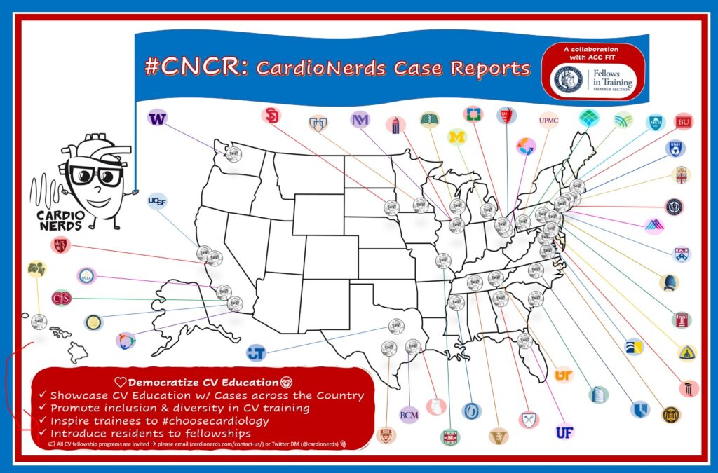
We are teaming up with the ACC FIT Section to use the #CNCR episodes to showcase CV education across the country in the era of virtual recruitment. As part of the recruitment series, each episode features fellows from a given program discussing and teaching about an interesting case as well as sharing what makes their hearts flutter about their fellowship training. The case discussion is followed by both an E-CPR segment and a message from the program director.
CardioNerds Case Reports Page
CardioNerds Episode Page
CardioNerds Academy
Subscribe to our newsletter- The Heartbeat
Support our educational mission by becoming a Patron!
Cardiology Programs Twitter Group created by Dr. Nosheen Reza
Patient Summary
A 64 y/o female with a history of venous and arterial embolism on anticoagulation, known PFO, and obesity presented after a fall. There was no loss consciousness, pre-syncopal symptoms, chest pain, aura, weakness, or palpitations. She had no recent preceding illness. When she arrived in the ED she was hypoxic to 87% on ambulation on room air and required 4L of nasal cannula O2 supplementation. The AGH CardioNerds were consulted!
On examination, the team noted that upon sitting up the patient would desaturate to the mid 80% but when lying down oxygen saturation would improve to 95%! Her ECG demonstrated a RBBB and no acute ST-T changes. TTE was obtained and showed normal LV and RV function and size, no valvular disease, and a likely PFO on an agitated saline study. Cardiac MRI revealed mild RV hypertrophy and Qp:Qs of 0.8 (right/pulm cardiac output < left/systemic cardiac output, indicating right to left shunting). RHC showed normal right-sided pressures. A right femoral vein bubble study was done showing torrential right to left shunting! She underwent PFO closure and her platypnea-orthodexia resolved; she was discharged on room air.
Case Media

Episode Schematics & Teaching

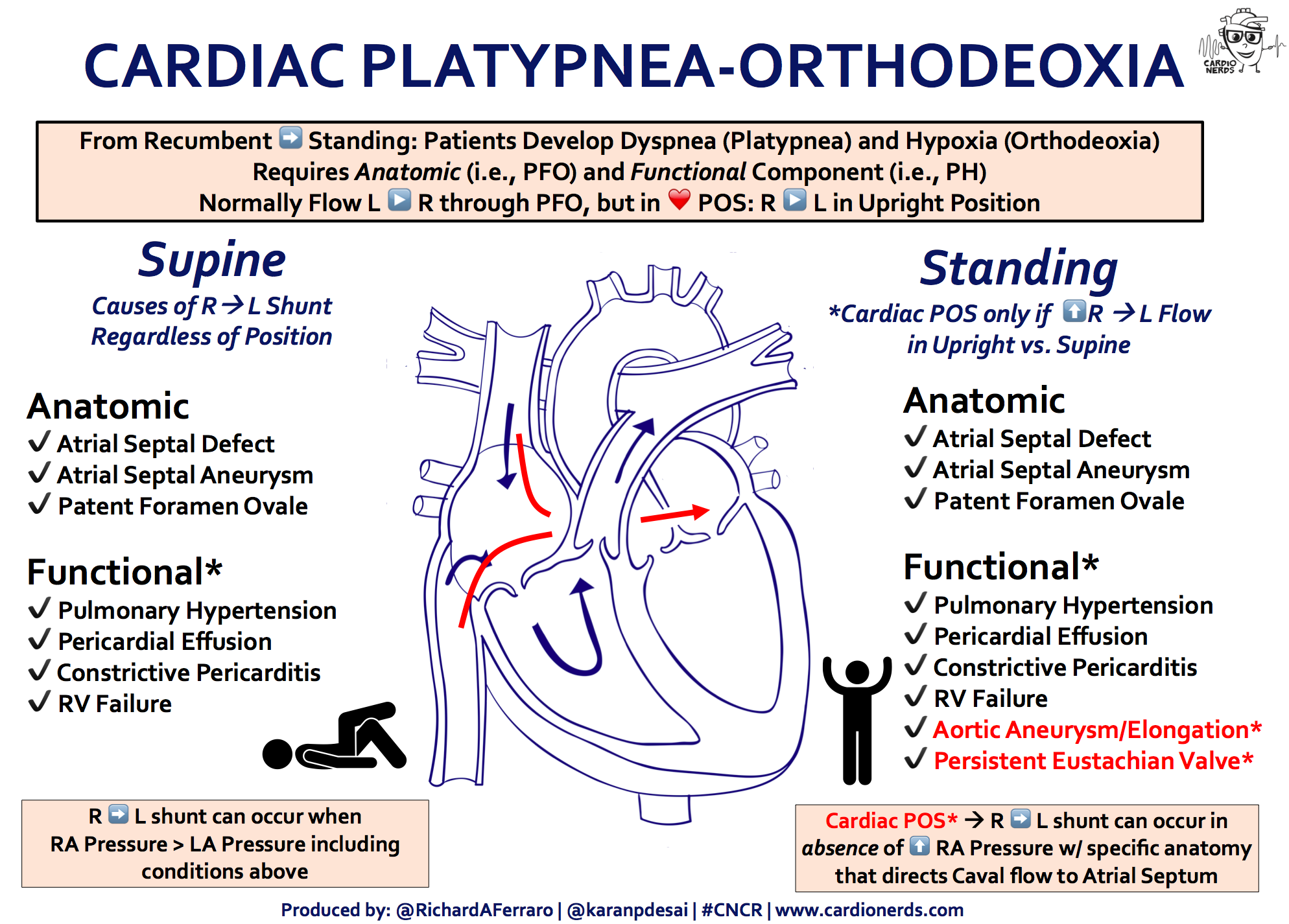
The CardioNerds 5! – 5 major takeaways from the #CNCR case
- Platypnea-orthodeoxia syndrome (POS) is when dyspnea (Platypnea) and hypoxia (Orthodeoxia) are present in the upright position, but improve upon lying flat. One cause is ARLIAS, or Acute Right-to-Left Inter-Atrial Shunting, which requires an anatomic component (I.e., ASD or PFO) and a functional component (I.e., PH, PE, RV failure) for Right-to-Left shunting to occur
- A PFO, or patent foramen ovale is a common congenital defect and typically will not lead to hypoxia unless there is right to left shunting. Typically flow is left to right due to an LA to RA pressure gradient and lower compliance of the RV.
- Remember when evaluating for a PFO with agitated saline, the timing of the appearance of bubbles is important! Early appearance of bubbles (seen in less than 5 beats of the cardiac cycle, think about intra-cardiac shunt. Delayed opacification (> 5 to 6 beats) occurs when the bubbles slowly build in the LV with each successive beat as they circulate to the LV and suggests extra-cardiac shunt.
- Cardiac MRI is helpful in quantifying the shunt fraction (Qp:Qs). This is the ratio of pulmonary flow (Qp) to systemic flow (Qs), where hemodynamically significant Left-to-Right shunt is > 1.5, especially when we see RA/RV dilation. Qp/Qs < 1.1 indicates net R to L shunt. Other ways to measure a Qp:Qs are echocardiogram (less accurate) and right heart catheterization.
- If there remains clinical concern for PFO with right to left shunting without evidence of a clear functional cause clinically or by catheterization (I.e., no evidence of high PA pressures, pericardial effusion, constrictive pericarditis), consider a right femoral vein bubble study. Upper extremity agitated saline enters the RA via the SVC; however, patients with prominent eustachian valve at the IVC can have blood flow directed towards the interatrial septum. This will be demonstrated on femoral vein bubble study!
Educational Video
References
- Cheng, T. O. (2002). Mechanisms of platypnea-orthodeoxia: what causes water to flow uphill. Circulation, 105(6), e47.
- Tobis, J. M., & Abudayyeh, I. (2016). Platypnea-orthodeoxia syndrome: an overlooked cause of hypoxemia.
- Sanikommu, V., Lasorda, D., & Poornima, I. (2009). Anatomical factors triggering platypnea‐orthodeoxia in adults. Clinical Cardiology: An International Indexed and Peer‐Reviewed Journal for Advances in the Treatment of Cardiovascular Disease, 32(11), E55-E57.
CardioNerds Case Reports: Recruitment Edition Series Production Team
-

Bibin Varghese, MD -

Rick Ferraro, MD -
Tommy Das, MD -

Eunice Dugan, MD -

Evelyn Song, MD -
Colin Blumenthal, MD -

Karan Desai, MD -

Amit Goyal, MD -

Daniel Ambinder, MD